Labelled Diagram Plant Cell Under Light Microscope / Draw And Label The Diagram Of A Typical Animal Plant Cell Both Under Light Microscope Brainly In : Here's a photo of a plant cell under an electron microscope.
byKim Keane-0
Labelled Diagram Plant Cell Under Light Microscope / Draw And Label The Diagram Of A Typical Animal Plant Cell Both Under Light Microscope Brainly In : Here's a photo of a plant cell under an electron microscope.. Juicy green plant cells under the microscope. Answer the following questions in your exercise book. Label the circle with the appropriate magnification. A cell is a very tiny structure which exists in living bodies. Let's go over the individual components of plant cells listed on a diagram such as the one above, and explore the roles that each of the organelles has.
When we look at cells under the microscope, our usual measurements fail to work. Elodea species or other freshwater aquarium plant. But at the same time it is interpretive. The animal cell and plant cell diagrams are easily colorable, allowing students to differentiate the different parts of the cell quickly. For example, iodine is often used to stain plant cells because it colours the starch.
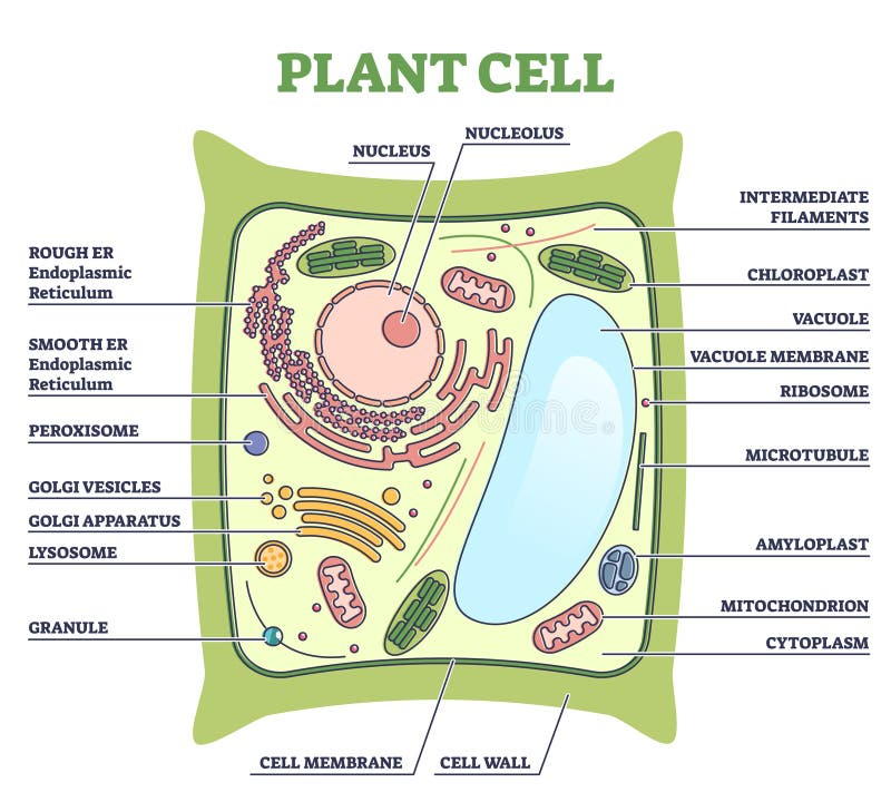
Animal Cell Definition Structure Parts Functions And Diagram from microbenotes.com A cell is a very tiny structure which exists in living bodies. Plant cells contain many organelles such as ribosomes, the nucleus, the plasma membrane, the cell wall, mitochondria, and chloroplasts. Juicy green plant cells under the microscope. 1 lab plant and animal cells, light microscopic analysis of leaf cross sections upper, structure of animal cell and plant cell under microscope, vacuole stock photos vacuole stock images alamy, cell structure teaching resources the science teacher. The plant cell is the basic structural and functional unit some of these differences can be clearly understood when the cells are examined under an electron microscope. Structure of animal cell and plant cell under microscope. Labelled diagrams of the four main cell types: Light photomicrograph of helianthus stem cross section seen through microscope.
Students will observe onion cells under a microscope.
Plant cells are the basic unit and building blocks of life in organisms of the kingdom plantae. Light microscopes (also known as optical microscopes) are the original microscopes. It is published by the american society of plant biologists. Plug in the microscope and turn on the light source. A cell is a very tiny structure which exists in living bodies. Label the circle with the appropriate magnification. Light microscopes use a number of lenses to produce an image that can be viewed directly at the eyepiece. Labeled diagram of plant cell, created with biorender.com. The typical characteristics that define the plant cell include cellulose, hemicellulose and pectin, plastids which play a major role in photosynthesis and storage of starch, large vacuoles responsible for regulating the cell turgor pressure. For example, iodine is often used to stain plant cells because it colours the starch. Animal cells under a light microscope. Light microscope v electron microscope • image of pollen grains under the light microscope • image plant cells under electron microscope what is a cell? Your plant cells under microscope stock images are ready.
The high resolving power makes the electron microscope a very important research tool in microbiology. Under the microscope, it shows many different parts. Stains interact with a specific part of the sample, turning it a different colour from its surroundings. Record the microscope images using labelled diagrams or produce digital images. Light microscopes (also known as optical microscopes) are the original microscopes.
Plant Cell Stock Illustrations 8 645 Plant Cell Stock Illustrations Vectors Clipart Dreamstime from thumbs.dreamstime.com (glow) cell ultrastructure • in the you must include… labelled diagrams of the four main cell types: Image:plant cell seen under electron microscope. Compound light microscope · explain why objects must be centered in the field of view before going from low to high power using. Observe the labeled diagram of plant. Label the circle with the appropriate magnification. Learn about the size and function of plant and animal cells for gcse biology, aqa. Robert hooke in 1665 first discovered plant cell. (refer to box 7.1 on p.
34 animal cell under electron microscope many new structures can be seen in the cytoplasm that are not seen using a light microscope.
Plant and animal cells lab objectives:. The compound microscope is a precision instrument. The high resolving power makes the electron microscope a very important research tool in microbiology. (glow) cell ultrastructure • in the you must include… labelled diagrams of the four main cell types: Does anyone have a decent labelled diagram of a plant cell under an electron microscope? Plant cells contain many organelles such as ribosomes, the nucleus, the plasma membrane, the cell wall, mitochondria, and chloroplasts. Labelled diagrams of the four main cell types: The typical characteristics that define the plant cell include cellulose, hemicellulose and pectin, plastids which play a major role in photosynthesis and storage of starch, large vacuoles responsible for regulating the cell turgor pressure. Light photomicrograph of helianthus stem cross section seen through microscope. The term 'cell' was coined to describe the small walled units that were observed in the sections they are just visible as small rods or spheres under light microscope. Plant cells are the basic unit and building blocks of life in organisms of the kingdom plantae. Structure of animal cell and plant cell under microscope. Organelles in a plant cell may not be present in an animal cells.
Structure of animal cell and plant cell under microscope. Plant cells contain many organelles such as ribosomes, the nucleus, the plasma membrane, the cell wall, mitochondria, and chloroplasts. Let's go over the individual components of plant cells listed on a diagram such as the one above, and explore the roles that each of the organelles has. Label the structures that you can see (e.g., cell wall repeat step 7 for higher settings of the microscope. Elodea species or other freshwater aquarium plant.
Plant Cell Structure Plant Cell Parts Organelles And Their Functions And Diagram Jotscroll from www.jotscroll.com Labeled diagram of plant cell, created with biorender.com. Elodea species or other freshwater aquarium plant. This shows a draw and label the structure of a generalized animal cell (i.e. Labelled diagrams of typical animal and plant cells with editable layers. Draw a diagram of one cheek cell and label its parts. Under the microscope, it shows many different parts. For example, iodine is often used to stain plant cells because it colours the starch. Observe the labeled diagram of plant.
Plug in the microscope and turn on the light source.
Be sure to only use the fine focus at the highest setting. This shows a draw and label the structure of a generalized animal cell (i.e. The compound microscope is a precision instrument. When we look at cells under the microscope, our usual measurements fail to work. When you look at animal or plant cells under the electron microscope, you can see a lot. 129 of your text book for precise instructions on. With light microscopy i can simply scrape some cells from my cheek smear them on a slide and look at them. Although plant cells differ greatly they all have similar eukaryotic the chloroplast is the area of the plant that is involved in the conversion of light energy into starch a plant contains specialised phloem cells, these enable the transport of signalling plant hormones and. Dreamstime is the world`s largest stock photography community. The animal cell and plant cell diagrams are easily colorable, allowing students to differentiate the different parts of the cell quickly. But at the same time it is interpretive. A cell is a very tiny structure which exists in living bodies. Record the microscope images using labelled diagrams or produce digital images.
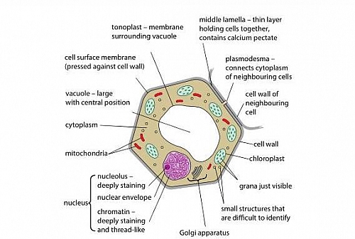
Although plant cells differ greatly they all have similar eukaryotic the chloroplast is the area of the plant that is involved in the conversion of light energy into starch a plant contains specialised phloem cells, these enable the transport of signalling plant hormones and plant cell under light microscope diagram. Plant and animal cells lab objectives:.


Post a Comment